

The essential answer
Cellular senescence is a complex state that cannot generally be identified by one universal marker. A senescence-associated signal can occur in other contexts, and different senescent populations need not share every feature. Interpret marker panels together with cell identity, persistence, and the biological question. A lower marker signal does not by itself demonstrate rejuvenation or improved function. In vitro evidence about a cell population remains distinct from claims about aging or treatment outcomes in humans.
How to reason about it
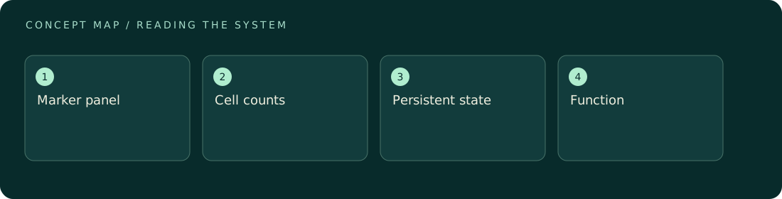
A marker is useful because it changes the plausibility of an interpretation, not because it creates a perfect label. Growth arrest, stress-related signals, morphology, and secretory features can provide complementary information, but their specificity depends on the system. The cited MICSE consensus paper documents the challenge of identifying senescence across contexts; it is guidance rather than a primary efficacy experiment. For original analysis, ask whether a marker shift reflects a state change within cells, a change in population composition, or loss of the cells that carried the marker. Those possibilities can yield the same average signal. A strong report makes its operational definition visible and explains conflicting endpoints. Avoid forcing every observation into a binary senescent-versus-young category when the measurements support only a narrower, context-specific characterization.
ILLUSTRATIVE · NOT A REPORTED RESULT
An example to work through
Assume a hypothetical culture contains 100 cells, of which 40 meet a predeclared marker criterion. Later, 20 of 80 remaining cells meet it. The fraction falls from 40% to 25%, but the counts alone do not show whether cells changed state or whether marker-positive cells were preferentially lost. Tracking function and population composition would be necessary to distinguish those explanations. This numerical example is a diagnostic illustration, not an intervention result, and it does not define a validated senescence threshold or a clinical endpoint.
Where interpretation fails
A decrease in one marker may be caused by altered expression, assay sensitivity, cell loss, or composition. Calling it age reversal skips those alternatives. Likewise, absence of a particular marker cannot rule out every senescence program in every cell type or tissue.
The next decision
State the operational criteria, use complementary evidence, and report cell counts alongside marker fractions. Keep discordant findings visible. If the data cannot distinguish state change from selective loss, describe the observed marker change without assigning rejuvenation or a beneficial human effect.
Sources & context
These sources support the concepts noted below. Our synthetic examples and research framing are educational interpretation; they are not findings from these organizations or validation of our ventures.
- Guidelines for Minimal Information on Cellular Senescence Experimentation in vivo ↗
Consensus guidance on marker limitations; used for interpretation boundaries, not efficacy.
Source check: Search-result abstract or official excerpt reviewed; full paper methods not audited
Educational research context, not a validated device specification or clinical protocol. Read our editorial and correction policy.