
The essential answer
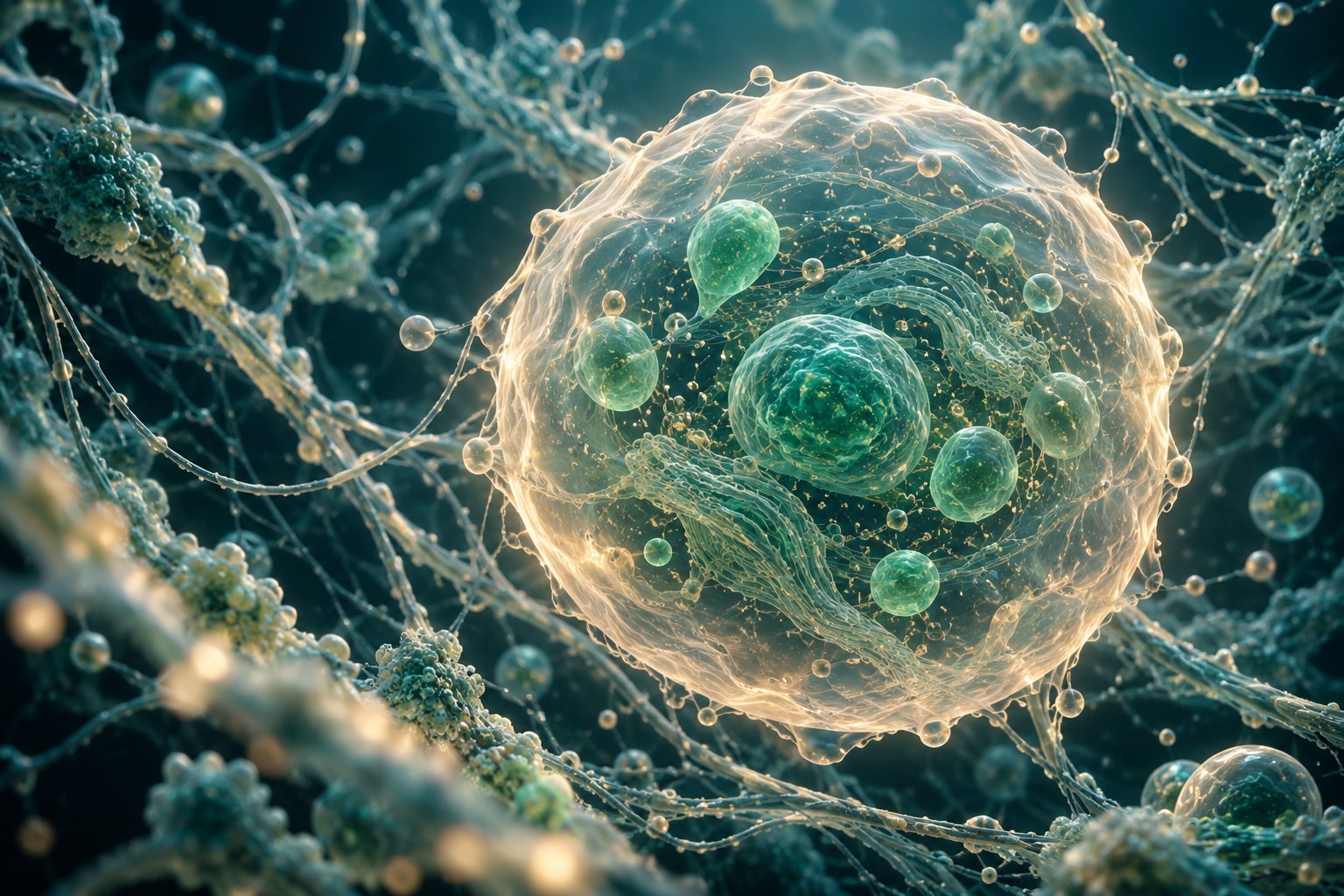
A microscope image can provide evidence about visible structure within the instrument's capabilities. It does not automatically establish that a cell-like object is living. NASA's microscopy research notes that size and shape alone can be difficult to use for distinguishing biological from nonbiological particles. The key learning habit is to separate observation from interpretation: first describe what the image shows, then state which additional evidence supports the proposed explanation. A convincing picture is still a measurement with limits.
How to reason about it
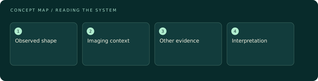
Images invite rapid recognition because humans are good at finding familiar shapes. That strength can also lead us to assign identity too quickly. A round boundary, an apparent division, or a moving spot can have more than one explanation. Scale, imaging mode, time sequence, and complementary information all affect what the image supports. A still image and a time series answer different questions; a rendered illustration and a measured micrograph are different kinds of evidence entirely. The educational task is not to distrust images, but to read them with the same precision applied to numbers. An annotation should distinguish an observed feature from a proposed interpretation, and processing or selection should be described when it materially affects what the viewer can conclude.
ILLUSTRATIVE · NOT A REPORTED RESULT
An example to work through
Assume two fictional images each show a round spot. The first is a measured image with a scale bar and acquisition context; the second is an artist's explanatory rendering. Their visual resemblance does not give them equal evidential roles. Now imagine that the measured spot moves between frames. Movement adds an observation, but without context it does not uniquely establish biological activity. The learner can write round feature and changed position before deciding whether stronger language is justified. This example uses no actual micrograph and makes no claim about a detected organism.
Where interpretation fails
Do not infer life from resemblance alone, and do not use a decorative rendering as if it were experimental evidence. Also avoid treating enhanced color or contrast as an intrinsic property without checking how the image was produced. Attractive presentation can clarify a finding, but cannot supply missing corroboration.
The next decision
Read the caption before interpreting the picture. Identify whether it is measured, modeled, or illustrative, then check scale, time context, and the claimed observation. Seek independent evidence for biological identity, and state morphology-based conclusions narrowly when competing explanations remain plausible.
Sources & context
These sources support the concepts noted below. Our synthetic examples and research framing are educational interpretation; they are not findings from these organizations or validation of our ventures.
- Microscopy Methods for Life Detection on Ocean Worlds ↗
NASA research states that size and shape alone can be insufficient for distinguishing biotic and abiotic particles.
Source check: Official research abstract opened; full report not reviewed.
Educational research context, not a validated device specification or clinical protocol. Read our editorial and correction policy.